
News


Biliary stent - the road to life again
ERCP, known as the jewel on the crown of digestive endoscopy, has recently introduced a set of duodenoscopy equipment and successfully performed endoscopic retrograde cholangiopancreatography (ERCP)+biliary stent implantation for an 85 year old patient, relieving biliary obstruction caused by malignant tumors of the common bile duct and establishing a pathway for the patient to regain their life.
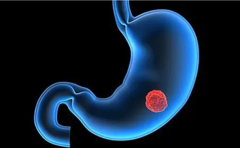
The patient came to our hospital for more than 10 days due to recurrent abdominal pain accompanied by yellowing of the skin and sclera, and worsening of yellow urine after 2 months. After hospitalization, it was found that the patient's abdomen was swollen, and a tumor about 3cm in size could be palpated in the upper right abdomen. The skin and sclera of the whole body were severely yellowed. MRI examination revealed significant dilation of the common hepatic duct, left and right hepatic ducts, and intrahepatic bile ducts. The gallbladder was full, suggesting malignant tumors of the common bile duct with biliary obstruction and intrahepatic metastasis. T3NxM1, obstructive jaundice, and the nature of the duodenal mass are unknown (duodenal cancer?). The conventional surgical treatment methods pose great risks to elderly patients, and considering the patient's physical condition and the complexity of the surgery, Qin Zeyu, the director of the Department of Gastroenterology at our hospital, organized expert consultations. After repeated discussions, it was decided to use ERCP technology to complete minimally invasive surgery, minimizing the trauma caused by the surgery to the patient.
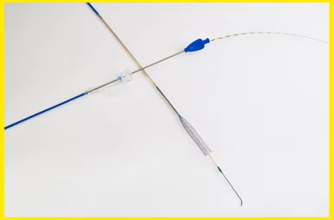
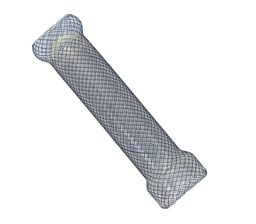
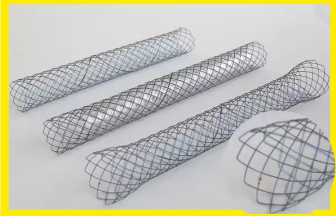
Surgical procedure: After successful anesthesia, the duodenoscope was smoothly inserted into the descending part of the duodenum, and the guide wire was smoothly inserted into the bile duct through the nipple opening. The angiography showed a significant eccentric stenosis of the left and right hepatic ducts towards the nipple side of the common hepatic duct, with a length of about 4cm. The left and right hepatic ducts and intrahepatic bile ducts were significantly dilated. First, a small incision of the nipple was made, and then a 6mm diameter expanding balloon was used to expand the nipple and narrow segment. Then, a 6X80mm non covered metal biliary stent was implanted. After the stent was released, a large amount of deep yellow bile gushed out, and the outer opening of the stent was located outside the nipple opening. The surgery went smoothly, and the patient's obstruction was relieved after surgery. The yellow staining gradually subsided, and they have now recovered and been discharged from the hospital.